3D Microanatomical Atlas
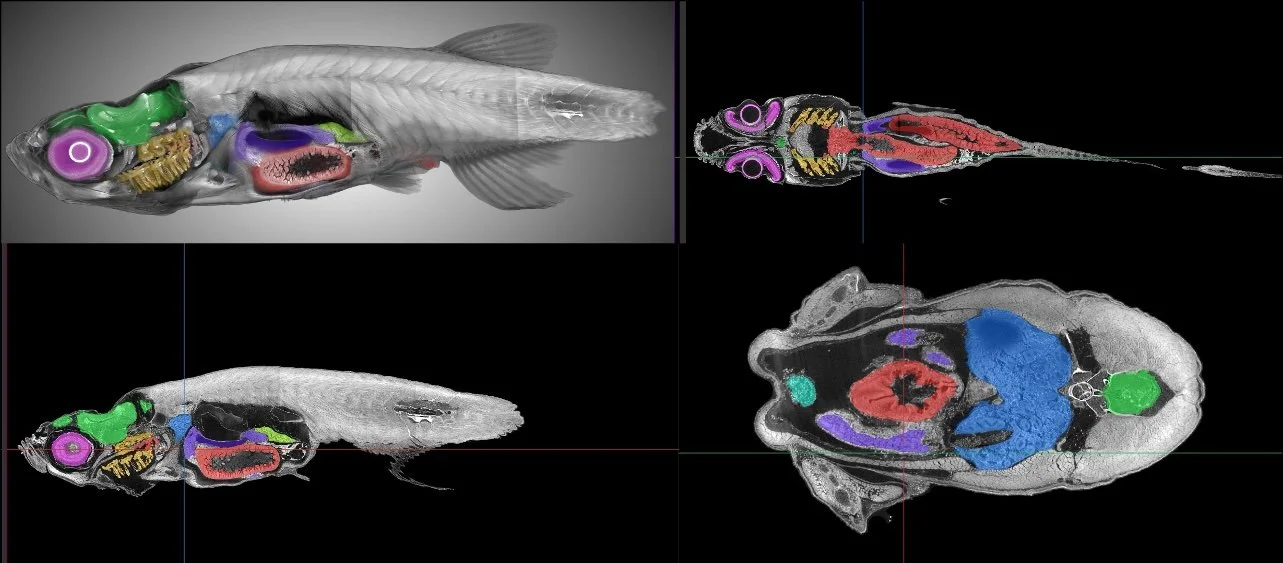
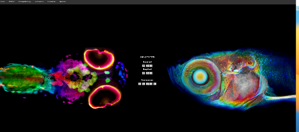
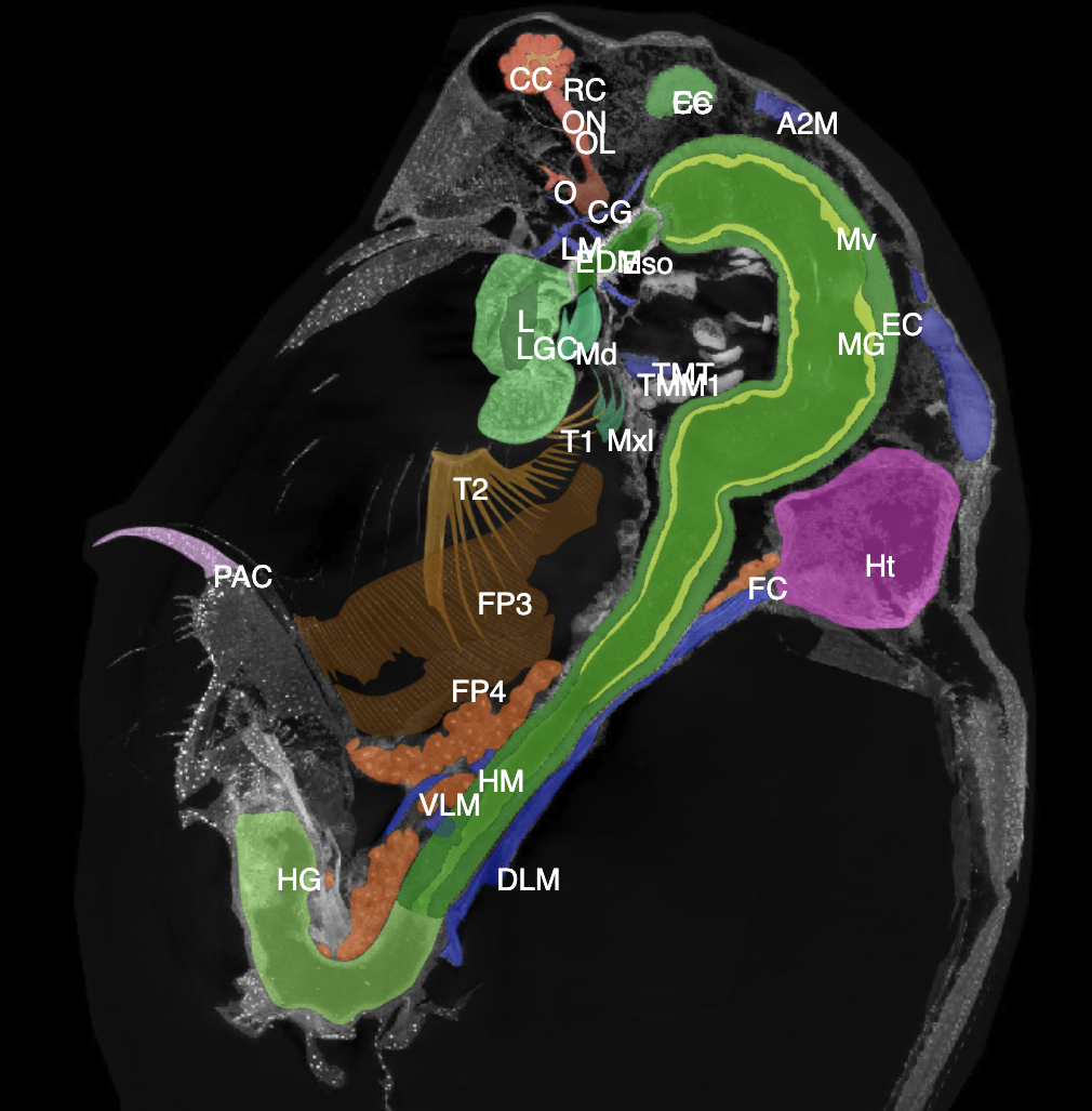
Whole-organism phenotyping is vital for understanding how genetic and environmental factors determine normal and disease phenotypes, and atlas resources that integrate unbiased “-omics” and tissue morphology would facilitate this goal. The pathology of cells and their arrangements remain fundamental endpoints in a wide variety of fields that require the study of gene function and organismal toxicity. To achieve more comprehensive insight, there is a compelling need to integrate multi-imaging technologies and molecular “-omics” data on a single atlas that enables unbiased phenotypic interrogation across all cell types. While traditional 2D histology provides subcellular detail, its inherent limitations includes its destructive, two-dimensional perspective, and prone to sampling artifacts, which restrict insights into the 3D structure and organization of cells within tissues. To overcome these limitations, we propose to add a microCT component to atlases that will add 3D representations of cells and organs, allowing researchers to interrogate the spatial relationships between the structures and digital exploration of microanatomy in virtual environments. MicroCT images will serve an integrative role, anchoring histology, fluorescence and ultrastructural imaging, and large-scale “-omic” data across length scales. We imaged Daphnia, zebrafish, and Drosophila at an unprecedented combination of 5 mm field-of-view (FOV) with 0.5 μm isotropic resolution at Lawrence Berkeley National Laboratory. Data are presented using the 4-planar viewer (sagittal, transverse, coronal and 3D), Neuroglancer. Segmentation of organ of interest can be done and incorporated onto the 3D file for visualization. By incorporating molecular and morphological data within a unified, organism-wide context, this 3D atlas platform enables multi-scale interrogation of how genetic variation and environmental perturbations drive phenotype. The tools of the 3D microanatomical atlas resource are being created as a foundation for future application across model organisms and human atlases to anchor research and educational efforts to increase understanding of biological structure, disease, and development.

